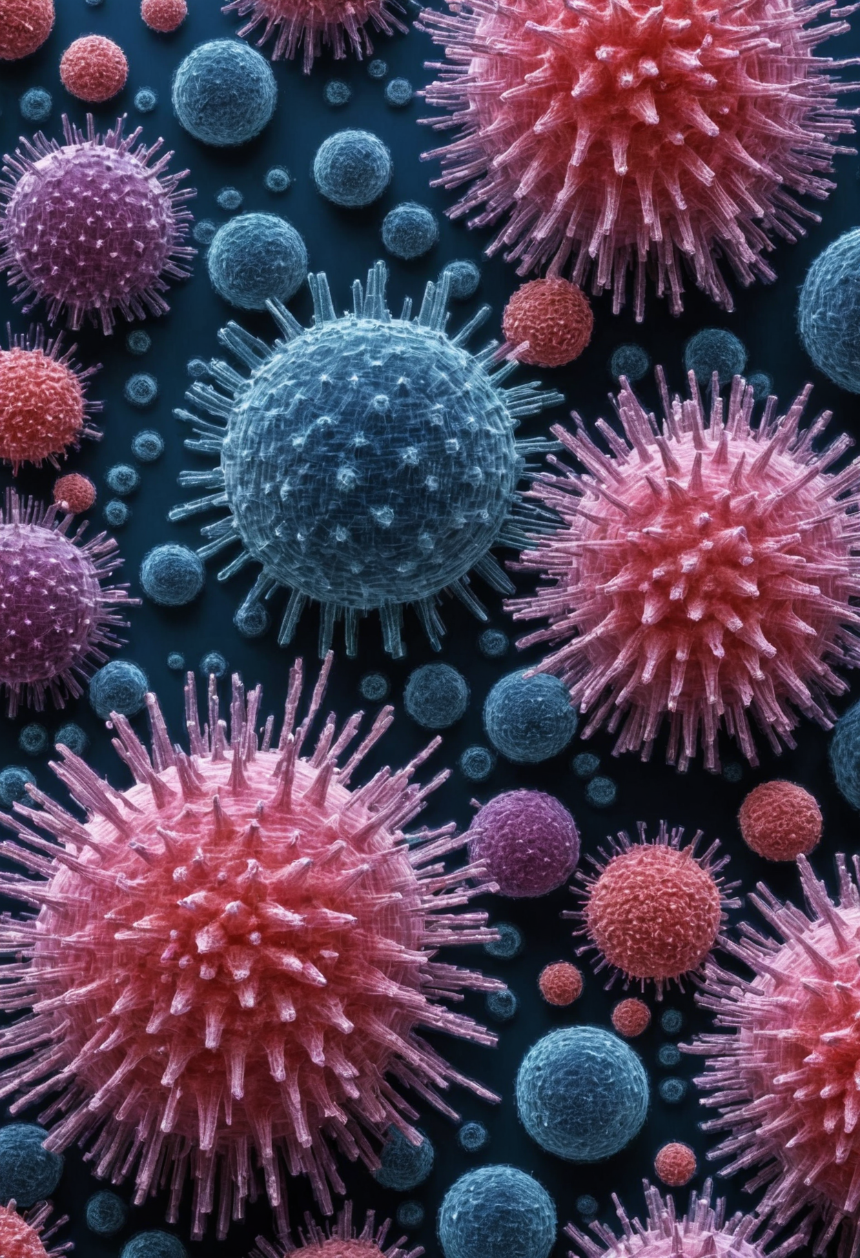
The image should represent a microscopic view of cells, likely captured through electron microscopy, a technique often used in scientific and medical research. The key elements of the image are spherical entities with a bumpy surface texture, colored blue, that are attached to a larger, irregularly shaped pink structure with filament-like projections. This pink structure should be the central focus of the image, with the blue cells attached to it in various places. The pink structure should appear slightly larger and more irregular in shape, suggesting a pathogen or cancerous cell. The blue cells should have a distinct spherical shape and a bumpy texture, representing immune cells. The image should be designed with a high level of detail and contrast, emphasizing the intricate relationships and interactions between the cells. This image is intended for medical professionals, researchers, and students in the field of biology or medicine, and should therefore be as scientifically accurate as possible. Given the complexity and scientific nature of the image, it is recommended for detailed analysis and expert interpretation. Comparative analysis with similar medical images could help viewers understand the type of cells and the specific biological process depicted in the image.
worst quality, low quality, low contrast, blurry, low quality, medium quality, watermark, username, signature, text, bad anatomy, bad hands, text, error, missing fingers, extra digit, fewer digits, cropped, jpeg artifacts, bad feet, extra fingers, mutated hands, poorly drawn hands, bad proportions, extra limbs, disfigured, bad anatomy, gross proportions, malformed limbs, missing arms, missing legs, extra arms, extra legs, mutated hands, fused fingers, too many fingers, long neck